
This paper focuses on the role of interventional radiology embolisations in a sequence of sufferers presenting with iatrogenic vascular injuries of the lower limbs following orthopaedic interventions.Fourteen sufferers (imply age: 64 years, vary 23-90 years) have been retrospectively analysed.
Clinical presentation consisted of palpable pulsatile mass, ache, decreased lower limb movement, or seen haematoma; 11 sufferers had additionally anaemia (haemoglobin < 7 g/dl).
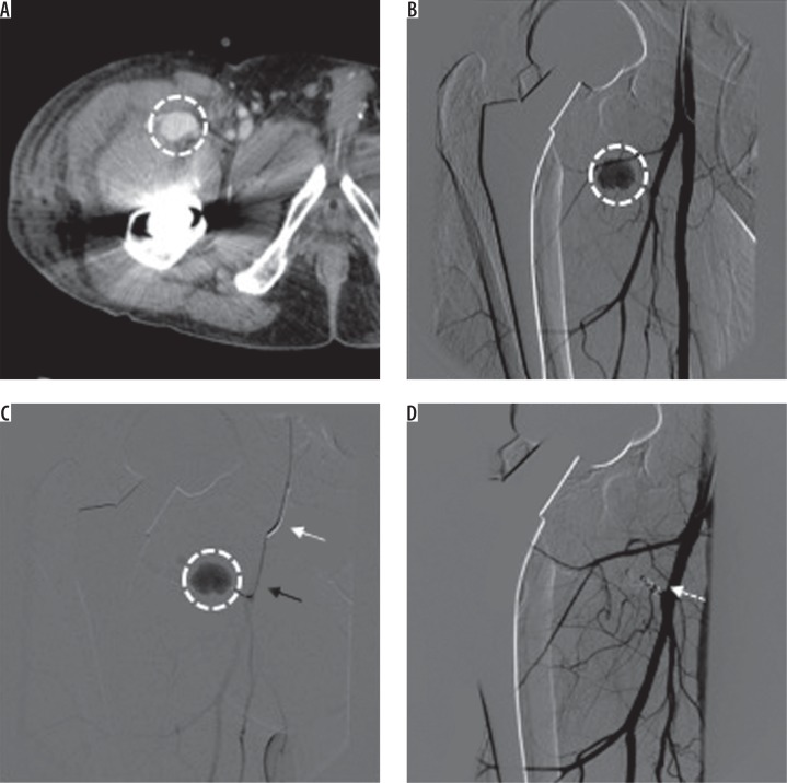
The time between orthopaedic surgical procedure and embolisation ranged between zero and 67 days (imply: 15 days). Injured arterial vessels have been as follows: inferior gluteal artery (2), superficial exterior pudendal artery (2), deep femoral artery
(1), lateral circumflex femoral artery (3), medial circumflex femoral artery (2), articular department of descending genicular artery (1), perforating femoral arteries (3), posterior tibial recurrent artery (1), and anterior tibial artery
(1). The typologies of vascular lesion have been: pseudoaneurysm 57%, bleeding with extraluminal distinction agent blush of the terminal arterial phase 36%, and laceration and bleeding with extraluminal distinction agent blush of the arterial predominant trunk 7%. Embolising brokers adopted have been microcoils 57%, glue 14%, microplug 7%, particles 14%, and coated stent 7%. In all instances medical and procedural technical successes have been obtained (100%).
For the administration of vascular injuries occurring after completely different orthopaedic interventions of the lower limbs, endovascular embolisations have confirmed to be secure and efficient; orthopaedic surgeons needs to be conscious of the assist that interventional radiology might present in the case of iatrogenic vascular problems.
The historical past of bone marrow in orthopaedic surgical procedure (half I trauma): trepanning, bone marrow injection in injury management resuscitation, and bone marrow aspiration to heal fractures.
One of the oldest procedures carried out by man is trepanning of the bone and but it was solely in the final 40 years that bone marrow aspiration has been used to deal with nonunion problems.
These advances have been attainable because of enhancements in devices and in strategies to make holes in the bone, an historical past that started with cranium trephinations round 8000-10,000 years in the past, and continued with sternum bone marrow injection for trauma resuscitation in the starting of the twentieth century; this process had improved at the starting of the twenty-first century to permit pelvis bone marrow aspiration for the treatment of nonunion.
Trephined skulls from antiquity have been discovered in many components of world, exhibiting that trephining was historical and widespread. Beginning with Neolithic interval and the pre-Columbian Andean civilizations, the authors have traced the growth of this surgical talent by describing the varied surgical instruments used to carry out holes in the cranium. These instruments (trephines or trepan) have been proposed at the finish of the nineteenth century to check the bone marrow.
At the starting of the twentieth century, the sternum grew to become the middle of curiosity for the “in vivo” research of the bone marrow and the fluid injection in the sternum’s bone marrow was described for resuscitation from shock throughout the World War II. With the introduction of plastic catheters and improved cannulation strategies, the want for intraosseous infusion as a substitute route for intravenous entry diminished and generally deserted.
However, throughout the mid-1980s, James Orlowski allowed renaissance of the use of intraosseous infusion for paediatric resuscitation. Since then, this method has develop into widespread and is now acknowledged as a substitute for intravenous entry in grownup emergencies; notably, the intraosseous entry has obtained class IIA advice from the Advanced Trauma Life Support program supported by the American College of Surgeons Committee on Trauma and bone marrow infusion is now really helpful for “Damage Control” resuscitation.
Although the pelvis bone incorporates half of the physique’s marrow quantity, it was solely in 1950 that the pelvis was proposed as a supply for bone marrow aspiration and bone marrow-derived mesenchymal stem cells to enhance therapeutic of fractures.It might be a few years earlier than doing holes in the bone as orthopaedic trauma process might be relegated to the annals of historical past.